Microscopic Anatomy Of The Eye
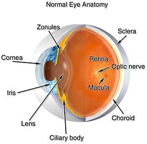
The retina is the light-sensitive tissue that lines the inside of the eye. The sclera is the layer at the top of the figure.
 Scanning Electron Microscopy Of The Human Cornea Eye Anatomy Anatomy Eye Facts
The microscopic anatomy of the eye of the Weddell seal was studied with various light and electron microscopic methods with a view to correlating morphological findings with the biology of this seal which is adapted to the extremes of the Antarctic environment and to extreme diving excursions into the lightless depths of the sea.

Microscopic anatomy of the eye. Learn vocabulary terms and more with flashcards games and other study tools. Click to see full answer Also question is what is microscopic anatomy. Microscopic anatomy is the study of structures on a microscopic scale along with histology the study of tissues and embryology the study of an organism in its immature condition.
Arterial Blood Vessels of the Orbit. From violet to blue to green to yellow to orange to red. Visual acuitythe ability to discern letters numbers and objects from a distanceis essential for many tasks from recognizing a friend.
The microscopic anatomy of the eye of the Weddell seal was studied with various light and electron microscopic methods with a view to correlating morphological findings with the biology of this seal which is adapted to the extremes of the Antarctic environment and to extreme diving excursions into the lightless depths of the sea. Outside of all next the choroid is the pigmentary layer 10. Since so many microscope users rely upon direct observation it is important to understand the relationship between the microscope and the eye.
Microscopic Anatomy of the Eye. Immediately internal to the sclera is the choroid which is dark as it is highly pigmented. Start studying Microscopic Anatomy of the Eye.
Microscopic Anatomy of the eye. Gross anatomy is the study of structures large enough to be seen with the naked eye and also includes superficial anatomy or surface anatomy the study by sight of the external body features. Light entering the eye passes through the transparent retina until it reaches the rods and cones and excites these and they stimulate the nerves.
Gross macroscopic anatomy is the study of anatomical structures that can be seen by the naked eye such as the external and internal bodily organs. Our eyes are capable of distinguishing color in the visible portion of the spectrum. For ophthalmologists optometrists medical dental and optometry students eye-anatomy forms the basis for eye-pathology in diseases.
Microscopic Anatomy of the eye. The eye cannot perceive ultraviolet or infrared rays. The nerve-fibres are believed to be continuous with the rods and cones.
The object examined by the eyepiece is the magnified inverted real image projected by the objective. Microscopic eye movements vital for 2020 vision. When the human eye is placed above the eyepiece the lens and cornea of the eye look at this secondarily magnified virtual image and see this virtual image as if it were 10 inches from the eye near the base of the microscope.
JOURNAL OF MORPHOLOGY 248165174 2001 Microscopic Anatomy of the Eye of the Deep-Diving Antarctic Weddell Seal Leptonychotes weddellii Ulrich Welsch1 Sven Ramdohr2 Bernd Riedelsheimer1 Rudolf Hebel1 Regina Eisert3 and Joachim Plötz2 1 Anatomische Anstalt Ludwig-Maximilians Universität München Germany 2 Alfred-Wegener Institut für Polar- und Meeresforschung Bremerhaven Germany 3 Animal and Food Sciences Division Lincoln University of Canterbury New Zealand ABSTRACT The. Dry eye retinal detachment macular degeneration diabetic retinopathy eye-trauma etc. Anatomy of the eye includes lacrimal gland cornea conjunctiva uvea iris choroid ciliary body lens blood supply retina vitreous optic-nerve.
EyeEar Microscope Learn with flashcards games and more for free. 3B MICROanatomy Human Eye Model - 3B Smart Anatomy Eye Models The MICROanatomy Eye model illustrates the microscopic anatomical structure of the retina with choroid and sclera. The action of the light is probably in the first instance chemical.
Dog Cat Horse Rabbit Monkey. The retinal pigment epithelium appears as a thin dark layer that forms a boundary between the neural retina and. The optical elements within the eye focus an image onto the.
Microscopic anatomy is the study of tiny anatomical structures such as tissues and cells. At the nasal aspect of the eye the conjunctiva is modified by the formation of the caruncle and the plica semilunaris a remnant of the nictitating membrane of animals. The interior of the eye the vitreous chambe r is on the bottom.
The retina functions in a manner similar to film in a camera. The microscopic anatomy of the eye of the Weddell seal was studied with various light and electron microscopic methods with a view to correlating morphological findings with the biology of this seal which is adapted to the extremes of the Antarctic environment and to extreme diving excursions into the lightless depths of the sea. The bulbar conjunctiva is composed of epithelium and underlying stroma which is loosely connected to the sclera and episclera.
Bone Tissue Anatomy Google Search Human Anatomy And Physiology Anatomy And Physiology Anatomy Bones
 Rods And Cones Eye Anatomy Anatomia Do Olho Microscopios Corpo Humano
 Anatomy Of The Human Eye Eye Microscopic Section Labeled Human Anatomy And Physiology Eye Anatomy Anatomy
 900024 Jpg Visuals Unlimited Eye Photography Macro Photography Eyes Macro Photography
 Anatomy Of The Eye Human Eye Anatomy Eye Anatomy Eye Health Eye Doctor
 Eye Anatomy Functions And Structure Eye Anatomy Senses Anatomy
 A 3d Render Of Your Eye Under A Microscope Video Video Things Under A Microscope Eyeball Art Eye Art
 Pin On Aia Course Human Vision
 The Eye And Vision Eye Anatomy Eyeball Anatomy Eye Anatomy Diagram
 Eye Micro Anatomy Of Retina The Retina Notes Anatomy
 Medical School Histology Of The Human Eye Medical Laboratory Science Medical School Studying Histology Slides
 Pin By Keryee Morton On Unit 4 Anatomy Medical Anatomy The Retina Anatomy
 Anatomy Atlases Www Anatomyatlases Org Plate 16 306 Retina Microscopy Nerve Fiber Microscopy Optometry
 The Eye And Vision Arteries And Veins Tears In Eyes Sweat Gland
 Erinbiol3500 Optic Nerve Histology Slides Anatomy And Physiology
 Normal Microscopic Section Of The Eye Lid Eye Pathology Online Pathology Glands Anatomy
 The Eye And Vision Rectus Muscle Anatomy And Physiology Autonomic Nervous System
 Medical School Medical Laboratory Science Human Anatomy And Physiology Medical School Stuff
 Human Eye Eye Anatomy Biology Diagrams Eye Study
Post a Comment for "Microscopic Anatomy Of The Eye"